MR Elastography
An advanced medical imaging technique that non-invasively quantifies tissue elasticity.
MR Elastography: Diagnostic Innovation Through Tissue Elasticity Imaging
Conventional medical imaging has been able to capture morphological changes in tissues but has faced significant challenges in quantitatively evaluating their physical properties, particularly “stiffness.” This limitation has been especially problematic in assessing the progression of hepatic fibrosis and qualitative diagnosis of brain tumors, where clinicians have had to rely on invasive biopsy procedures.
MR Elastography was developed as an innovative solution to address these challenges (Muthupillai et al., 1995). By applying external mechanical vibrations and capturing their propagation using high-speed MRI, this technology enables non-invasive and quantitative measurement of tissue elasticity. This breakthrough technique has enabled a wide range of clinical applications, including early detection of liver cirrhosis, differentiation between benign and malignant brain tumors, and evaluation of musculoskeletal disorders, simultaneously reducing patient burden while improving diagnostic accuracy.
By augmenting conventional morphological imaging with physical property information, MR Elastography represents a next-generation medical imaging technology that provides more comprehensive and precise diagnostics, with promising prospects for widespread clinical adoption.


I participated in a comprehensive research and development project at Chiba University and the National Institute of Radiological Sciences (NIRS) aimed at expanding the clinical applications of MRE technology and improving diagnostic accuracy. This research adopted innovative approaches from both hardware and software perspectives to address the technical limitations and clinical application challenges inherent in conventional apporaches.
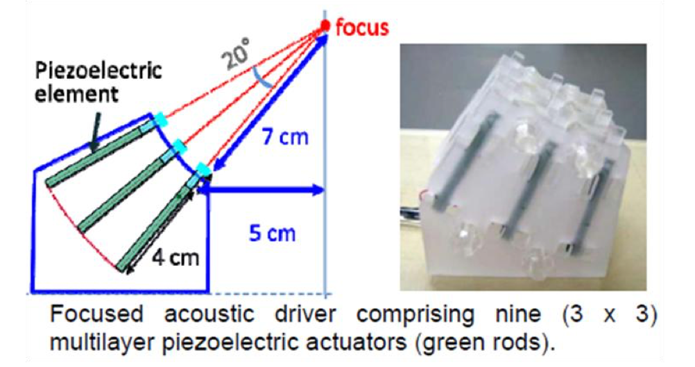
Effective MRE Measurement
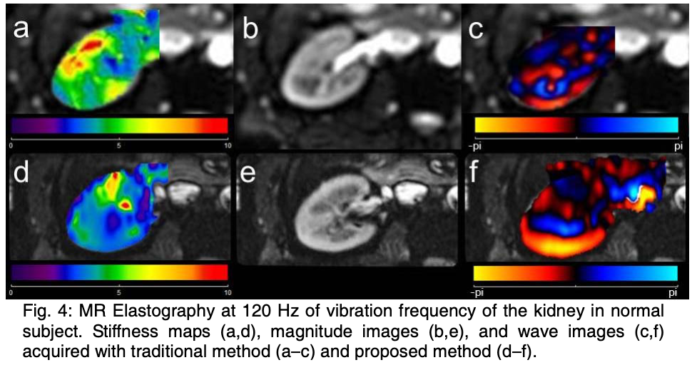
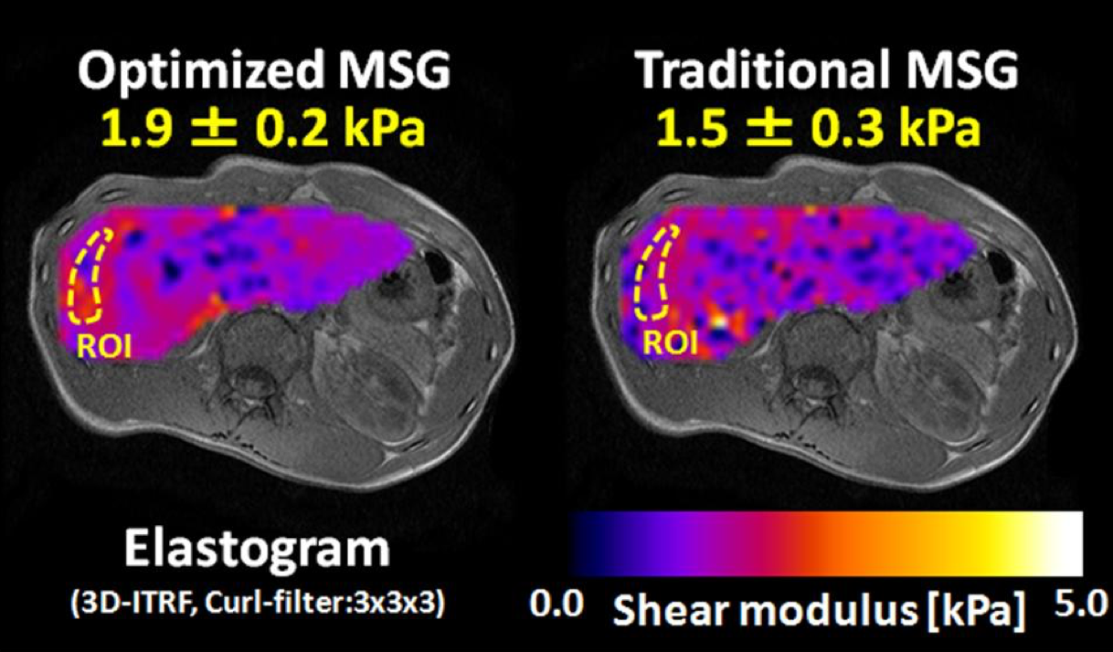
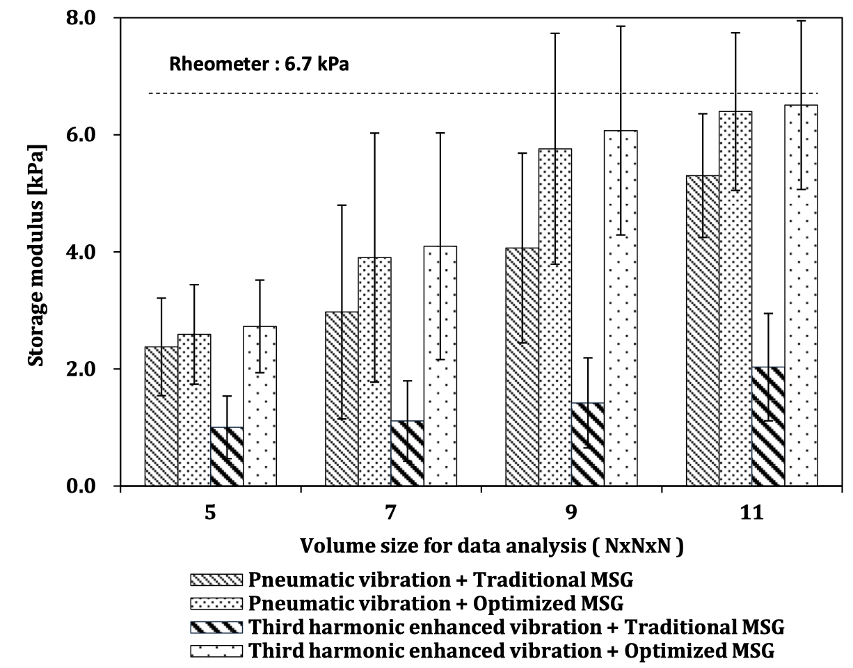
We introduced a novel multi-point convergent external vibration system to replace conventional single-point vibration methods, optimizing vibration propagation characteristics within tissues. Simultaneously, we implemented a transition from conventional differential-type calculation methods to integral-type algorithms for elasticity estimation (Suga et al., 2009), achieving significant improvements in noise tolerance and computational stability. In this project, I served a central role in MRI pulse sequence design and signal processing algorithm development.
Through gradient magnetic field control and RF pulse design in MRI imaging, I implemented selective extraction functionality for specific vibration frequency components (Suga et al., 2009; Suga et al., 2010; Ozawa et al., 2010; Kobayashi et al., 2010; Ozawa et al., 2010; Ozawa et al., 2011). I developed MR pulse sequence to precisely filter target frequency due to diverse biological vibration components and mechanical limitation, achieving substantial improvements in signal-to-noise ratio (SNR). Additionally, I constructed a real-time motion detection and correction system to eliminate artifacts caused by involuntary body movements during imaging, enhancing practical applicability in clinical environments. (Hirano et al., 2008; Arai et al., 2009; Arai et al., 2010; Arai et al., 2011; Suga et al., 2010; Arai et al., 2011; Arai et al., 2011)
Table Resonance Elastography in MR
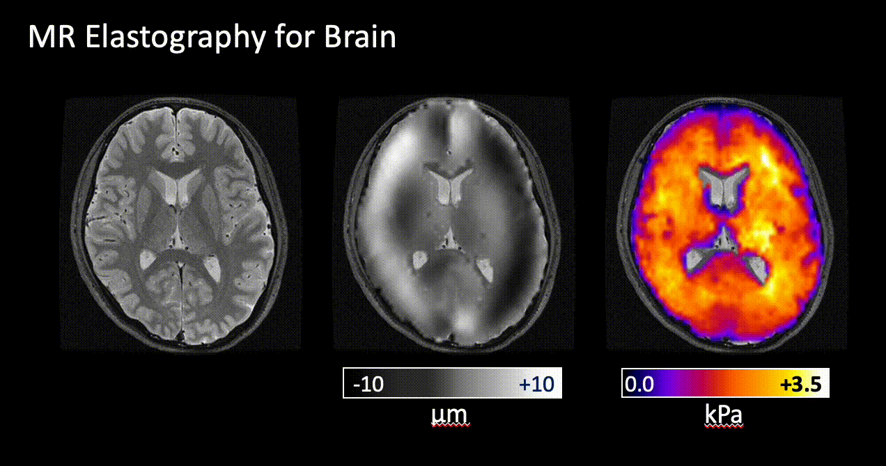
As one of groundbreaking achievements, I contributed to an external vibration device-free MRE imaging method utilizing the inherent physical characteristics of MR systems (Gallichan et al., 2009). By precisely controlling and utilizing minute vibrations generated during MR scanner coil operations, I developed a revolutionary approach that eliminates the need for conventional large external devices. This technology holds particularly innovative significance in the neurological field. It fundamentally solved the problem of direct vibration application to brain tissue, which was previously difficult due to physical constraints imposed by the skull, achieving the non-invasive brain elasticity measurement. (Arai et al., 2010; Ikeda et al., 2011)

MRE Under Free-Breathing
To overcome respiratory motion artifacts, the major technical challenge in abdominal organ MRE imaging, We developed a system integrating advanced image analysis and machine learning technologies. We constructed proprietary tracking algorithms that monitor diaphragmatic motion in real-time from MR signals and established technology for highly accurate identification of expiratory and inspiratory phases using random forest-based machine learning models. This technology eliminated the constraints of breath-hold imaging previously considered essential, enabling high-quality MRE examinations for patient populations where breath-holding is difficult, including elderly patients and those with chronic respiratory diseases. (Ozaki et al., 2014)

References
2014
2011
- Evaluation of the Effectiveness of a Multipoint Vibration System for MRE on a Living BodyIn The 60th National Congress of Theoretical and Applied Mechanics, Mar 2011
- Investigation of Imaging Methods in MR Elastography of the Human Abdomen - Comparison of Echo-planar and Gradient Echo Methods -In The 60th National Congress of Theoretical and Applied Mechanics, Mar 2011
- Preliminary Study of MR Elastography Using Gantry Vibration - Estimation of Optimum Imaging Parameters by Measuring Amplitude Characteristics -In The 60th National Congress of Theoretical and Applied Mechanics, Mar 2011
2010
- Examination of Vibration Vibration Apparatus for Improving Accuracy of Viscoelastic Modulus Estimation in MREIn The 59th National Congress of Theoretical and Applied Mechanics, Jun 2010
- Examination of Vibratory Apparatus for MRE Using Elastic Wave InterferenceIn The 59th National Congress of Theoretical and Applied Mechanics, Jun 2010
- Improvement of elastic modulus estimation accuracy of MR Elastography by multipoint excitationIn The 38th Annual Meeting of the Japanese Society for Magnetic Resonance in Medicine, Oct 2010
- Suppression of harmonic distortion on elastic wave images by optimizing pulse sequences for MR elastographyIn The symoposium for The Japanese Society for Medical and Biological Engineering 2010, Sep 2010
- Estimation Method for Viscoelastic Modulus Distribution Considering Compressibility of Living BodyIn The 59th National Congress of Theoretical and Applied Mechanics, Jun 2010
-
 Preliminary study of viscoelastic modulus distribution measurement of the brain by MR elastography using gantry vibrationIn The 38th Annual Meeting of the Japanese Society for Magnetic Resonance in Medicine, Oct 2010
2009
- Development of an External Focus Vibration Apparatus for MR Elastography to Improve Quantification and Spatial Resolution of Viscoelastic Modulus Distribution in VivoIn The 37th Annual Meeting of the Japanese Society for Magnetic Resonance in Medicine, Oct 2009
- Optimization of Pulse Sequences for MR Elastography for Single Frequency Elastic Wave Image AcquisitionIn The 37th Annual Meeting of the Japanese Society for Magnetic Resonance in Medicine, Oct 2009
2008
- Investigation of Pulse Sequence Optimization in MR ElastographyIn The 36th Annual Meeting of the Japanese Society for Magnetic Resonance in Medicine, Sep 2008